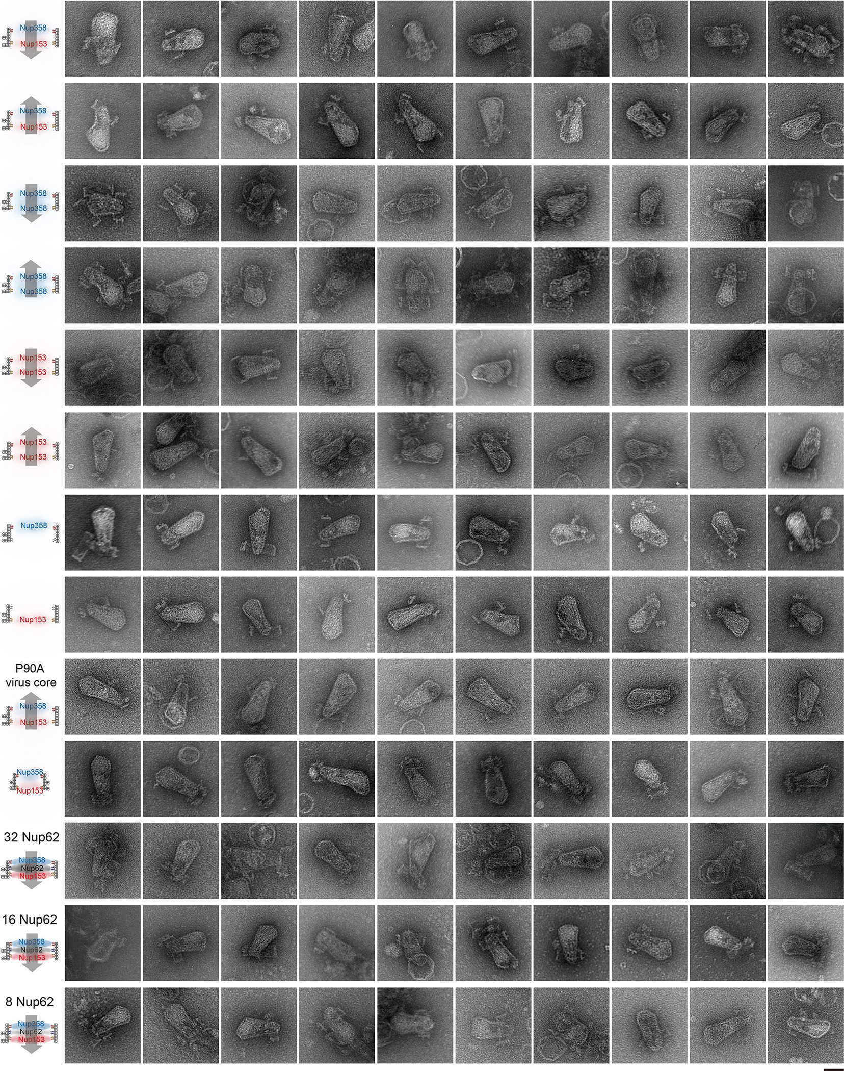
Extended Data Fig. 10 |. Galleries of representative negative-stain electron micrographs of HIV-1 core-NuPOD interactions.

The purified virus cores bound to NuPODs loaded with different nups, with schematics on the left. All negative-stain EM experiments were repeated three times with similar results. Scale bar, 50 nm. More high-magnification images of NuPOD-virus core interactions are included in Supplementary Fig. 1–7.
