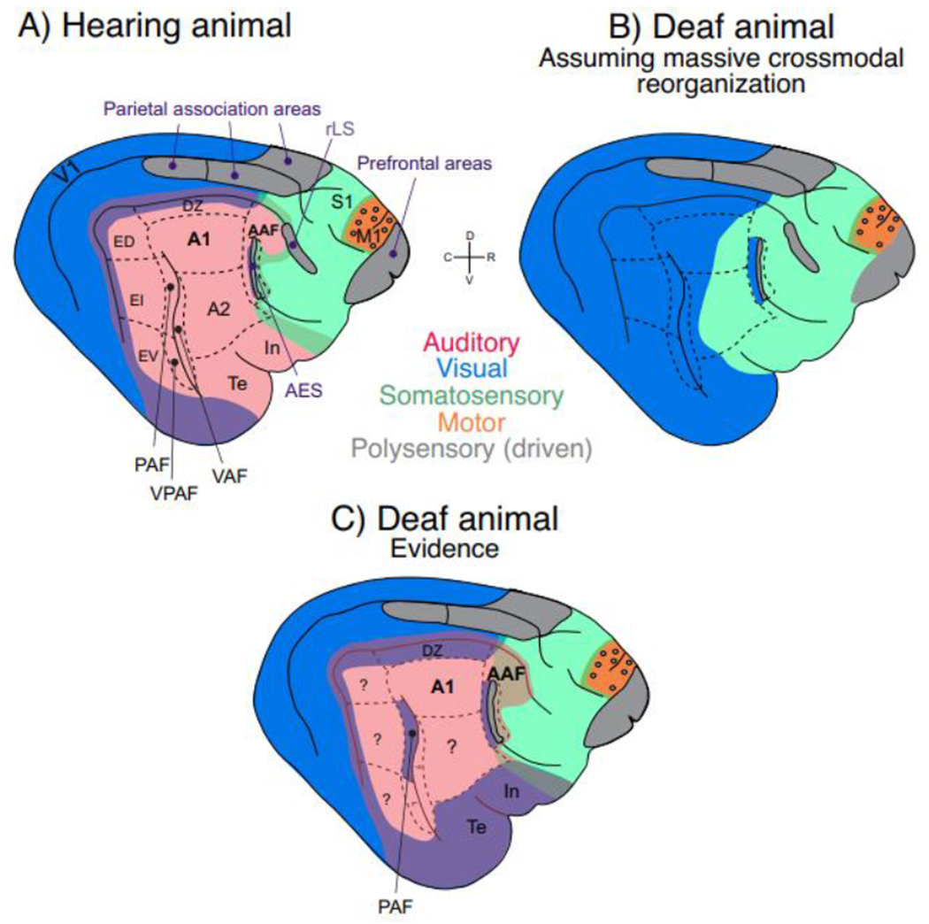
Fig. 1: Schematic illustration of approximate locations of sensory brain areas in the cat and their reorganization in deafness.

Blue – visual; red – auditory; green – somatosensory; orange – motor; grey – association areas. Mixture of colors at sensory borders (appears as violet, light brown/golden and deeper green) depicts bimodal responsiveness (~ two colors). A) AES, the area of the anterior ectosylvian sulcus, together with area In correspond to human insular cortex [4]. The ectosylvian sulcus (divided into dorsal area, ED, intermediate area, EI, and ventral area, EV) correspond to human superior temporal gyrus [5]. Feline prefrontal cortex is likely multisensory based on tracer studies [6]. Rostral lateral sulcus (rLS) and/or anterior ectosylvian sulcus (AES) allow multisensory integration in the superior colliculus [7]. “Parietal association cortex” are two separate areas within Brodmann area 7 [8,9]. Visual responsiveness was observed in the posterior belt in ED [5], but also in DZ. Auditory and visual responses have been described around the cruciate sulcus in M1. In the pericruciate association cortex visual responses and auditory responses were reported, here shown as grey circles within the motor cortex (orange). Cingulate cortex on the medial hemisphere and ventral limbic areas (entorhinal cortex and parahippocampal gyri) are not shown. B) A putative cortical map under the assumption of massive crossmodal reorganization in congenital deafness. C) An approximate representation of actual cortical organization in congenitally deaf cats according to the evidence as reviewed in the text.
