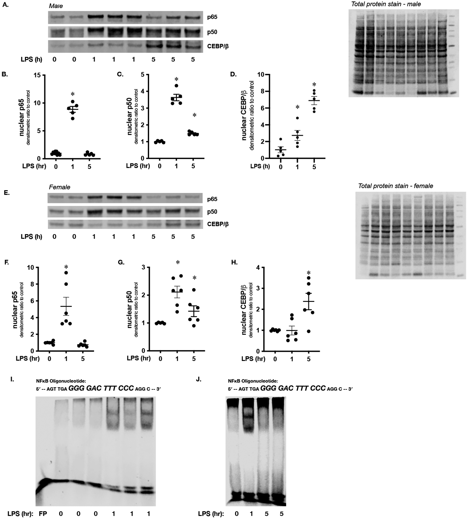
Figure 5. Sublethal endotoxemia induces hepatic NFκB and CEBP/β nuclear translocation.

(A) Representative Western blot of p65, p50 and CEBP/β on hepatic nuclear extracts isolated from endotoxemic (LPS 5 mg/kg, IP; 1 and 5 hours) male mice. Total protein stain as loading control. (B-D) Densitometric analysis of nuclear (B) p65, (C) p50 and (D) CEBP/β. Data normalized to total protein and expressed as mean ± SEM (N = 4–5 per time point). *p < 0.05 vs. unexposed control. (E) Representative Western blot of p65, p50 and CEBP/β on hepatic nuclear extracts isolated from female mice exposed to endotoxemia (LPS 5 mg/kg, IP; 1 and 5 hours). Total protein stain as loading control. (F-H) Densitometric analysis of nuclear (F) p65, (G) p50 and (H) CEBP/β. Data normalized to total protein and expressed as mean ± SEM (N = 4–5 per time point). *p < 0.05 vs. unexposed control. (I) Representative EMSA using an IR labeled oligonucleotide containing the NFκB consensus sequence (5’-AGTTGAGGGGACTTTCCCAGGC-3’) and hepatic nuclear extracts isolated from mice exposed to endotoxemia (LPS 5 mg/kg, IP; 1 hour). FP = free probe (J) Representative EMSA using an IR labeled oligonucleotide containing the NFκB consensus sequence (5’-AGTTGAGGGGACTTTCCCAGGC-3’) and hepatic nuclear extracts isolated from endotoxemic (LPS 5 mg/kg, IP; 1 and 5 hours) mice.
