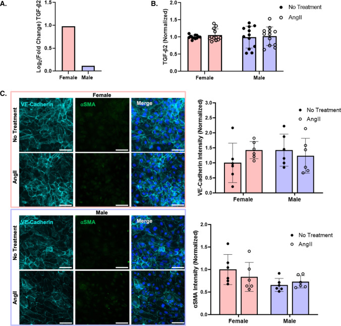
Fig. 2.
AngII increased TGFβ2 mRNA expression in female as compared to male HUVEC, but TGFβ2 protein and EndMT did not change with AngII or sex. A RNA-seq of changes in TGFβ2 gene expression following 24 h of 1 µM AngII treatment in pooled male and female HUVEC (n = 3 samples per condition). B TGFβ2 protein after 24 h of 1 µM AngII treatment in pooled male and female HUVEC, as measured by ELISA (n = 12 samples; 3 pooled HUVEC samples per condition in each experiment, with the experiment repeated 4 times). C Representative fluorescent microscopy images of endothelial (VE-cadherin, light blue) and mesenchymal (αSMA, green) markers, with Hoescht-labeled nuclei (dark blue) in female and male HUVEC following 24 h of 1 µM AngII treatment, with quantification (n = 6 samples; 3 pooled HUVEC samples per condition in each experiment, with the experiment repeated 2 times). Scale = 50 µm. TGFβ2 protein and fluorescence intensity were normalized to untreated female cells

