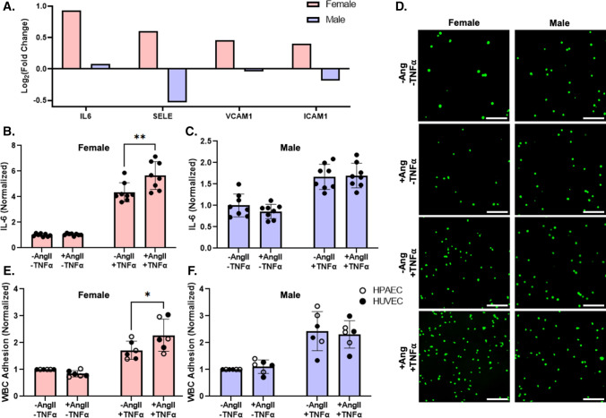
Fig. 3.
Female EC had higher IL-6 mRNA expression than male HUVEC following AngII treatment but required concurrent TNFα exposure to show higher IL-6 protein and WBC adhesion. A IL-6, SELE, VCAM1, and ICAM1 gene expression from RNA sequencing in pooled female and male HUVEC following 24 h of 1 µM AngII treatment (n = 3 samples per condition). B Female and C male pooled HUVEC IL-6 protein release following 24-h treatment ± 1 µM AngII and ± 1 ng/mL TNFα, measured via ELISA (n = 8 samples; 4 samples per condition in each experiment, with the experiment repeated 2 times). D Representative WBC adhesion fluorescent microscopy images (calcein, green). E Female and F male pooled HUVEC, individual donor HUVEC, and individual donor HPAEC quantified WBC adhesion following 24-h treatment ± 1 µM AngII and ± 1 ng/mL TNFα (n = 6 donors per sex; each datapoint averaged from 3 samples per donor per condition). Scale = 100 µm. Data analyzed with two-way ANOVA. *p < 0.05, **p < 0.01. IL6 protein concentration and WBC adhesion normalized to untreated cells for each sex

