FIG. 2.
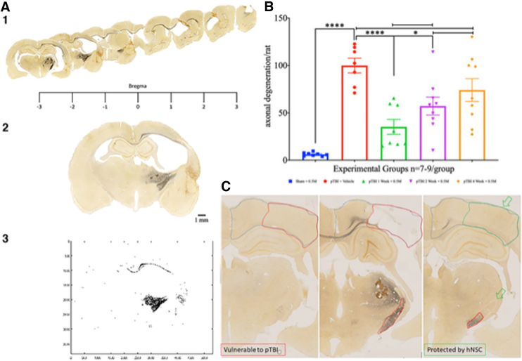
(A, 1–3) Rostrocaudal serial sections from a group 7 representative animal shows the extent of axonal degeneration (black, silver-stained regions) across the rodent brain. The rostral unilateral motor cortical injury turns bilateral in the corpus callosum, but is limited to the right hemisphere in caudal sections engulfing the corticofugal pathway, including the internal capsule (A-1). Representative silver-stained brain section (A-2) is thresholded and binarized (A-3) to digitally quantitate axonal damage. (B) Scatterplot of axonal degeneration (y-axis) shows a statistically significant difference between the sham (blue) versus pTBI (red; p < 0.0001), indicating a robust injury effect. Axonal degeneration increased proportional to interval length (green → purple → orange). Compared to group 7 (red), groups 2 (green) and 4 (purple) have significantly lower degeneration (p = 0.002 and 0.015, respectively). No differences were detectable upon comparison with group 6 (orange; p = 0.27). The one-way ANOVA had F3,36 = 15.61 and was followed by a Tukey's multiple comparisons post hoc test for pair-wise comparison. Within the injury + transplant, only group 2 was significantly better than group 6 (p = 0.024). (C) Axonal damage is absent in the section from the sham group (left image) compared to the group 7 section (pTBI + vehicle treated; middle image), which was replete with severe damage to the corpus callosum and internal capsule. Compared to group 7, delayed remote secondary damage, see the dorsal aspect of the internal capsule (green open arrow) was absent in group 2 animals (1-week interval; right image). All animals survived for 12 weeks after transplantation. Scale bar = 1 mm. ANOVA, analysis of variance; pTBI, penetrating traumatic brain injury.

