Fig. 5.
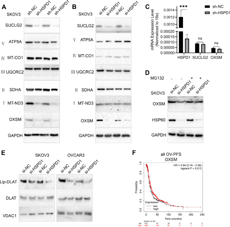
HSP60 knockdown disrupted mitochondrial protein-OXSM stability and lipoic acid synthesis. A, B Western blotting analysis was used to validate expression changes of individual mitochondrial proteins in HSP60 knockdown SKOV3 cells with siRNA and lentivirus shRNA, respectively. C Quantitative RT-PCR analysis was used to analyze the mRNA levels of HSPD1, SUCLG2 and OXSM. D SKOV3 cells were transfected with si-NC and si-HSPD1, 48 h later, cells were treated with or without 0.2 µM MG132 for 24 h prior to cell lysis. The level of OXSM protein was examined by western blotting. E Western blotting of lipoylated proteins (lip-DLAT) and total DLAT from extracts of OC cells with HSPD1 knockdown. F Kaplan-Meier survival analysis of progression-free survival between OXSM high and low transcript groups in all OC. The red line indicates patients with high OXSM levels and the black line indicates patients with low OXSM levels, log rank P = 0.01. ***P < 0.001

