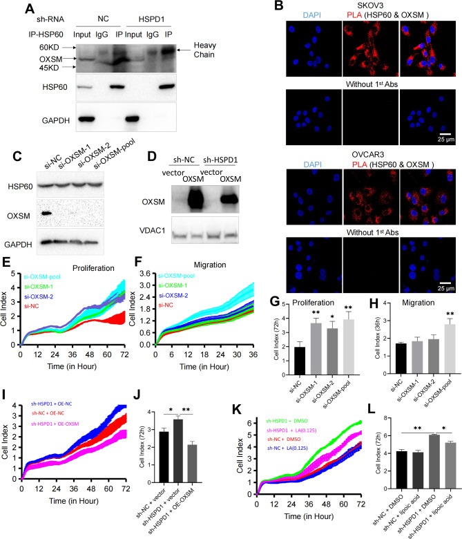
Fig. 6.
HSPD1 promotes ovarian cancer progression by maintaining function of OXSM. A Co-immunoprecipitation of OXSM with HSP60 in SKOV3 cells. B Examination of interaction of OXSM-HSP60 PLA signal in SKOV3 and OVCAR3 cells lines. Scale bar = 25 μm. C Western blotting analysis of OXSM expression in SKOV3 cells transfected with NC siRNA and OXSM siRNA. D Validation of the overexpression of OXSM in OC cells transfected with OXSM plasmid via western blotting. E, G Effects of OXSM knockdown with siRNA on the proliferation of SKOV3 analyzed by the RTCA assay. F, H Effects of OXSM knockdown on the migration of SKOV3 analyzed by the RTCA assay. I, J Cell proliferation was detected by RTCA assay after transfection with sh-HSPD1 and OE-OXSM. Quantification of the cell index after 72 h culture in the RTCA assay. K, L SKOV3 cells transfected with lentivirus sh-NC or sh-HSPD1 treated with lipoic acid (0 or 0.125 µM). Quantification of the cell index after 72 h culture in the RTCA assay. *P < 0.05, **P < 0.01

