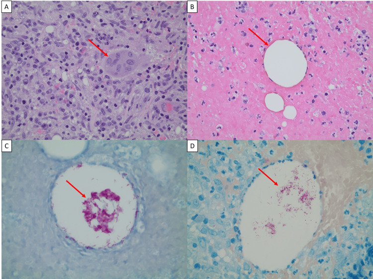
Figure 2. Histopathologic examination of excisional biopsy.
Surgical excision of lesion at the upper outer quadrant of the right breast. (A) Granulomatous inflammation with epithelioid histiocyte, multinucleated giant cells (red arrow), lymphocytes, and plasma cells (H&E, ×400). (B) Lipid-like vacuoles (red arrow) seen in the background of acute inflammation (H&E, ×400). (C) Clumps of intravacuolar acid-fast bacilli (red arrow, AFB stain, 600). (D) FITE stain highlights acid-fast microorganisms (red arrow, FITE stain, 600).

