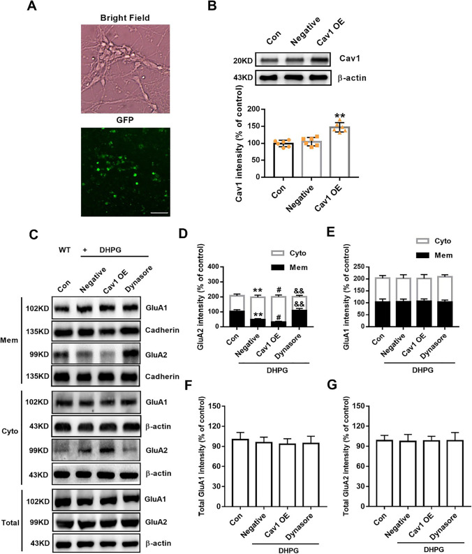
Fig. 5.
Overexpression of Cav1 regulates the GluA2 endocytosis induced by DHPG. A Bright-field image showing hippocampal neurons cultured in vitro for 10 days. GFP-positive neurons (green) indicate the successful infection of the lentivirus. Scale bar = 50 μm. B Cav1 expression was detected by Western blot after 4 days of infection. n = 6 dishes from three independent experiments; ** p < 0.01 versus negative control, one-way ANOVA with Tukey’s multiple comparisons test. C Representative western blots showed differential distribution of GluA1/2 on membranes and in the cytoplasm by DHPG-treated Cav1-OE WT neurons. D, E Quantitative analysis of cumulative western blot experiments. Immunoreactivities of GluA2 (D) and GluA1 (E) on membranes were normalized to cadherin, and the immunoreactivities in the cytoplasm were normalized to β-actin. n = 6 dishes from three independent experiments. F, G Quantitative analysis of cumulative western blot experiments. Immunoreactivities of total GluA1 (F) and GluA2 (G) were normalized to β-actin. n = 6 dishes from three independent experiments; ** p < 0.01 versus control; # p < 0.05 versus negative + DHPG treatment; && p < 0.01 versus Cav1-OE + DHPG treatment, one-way ANOVA with Tukey’s multiple comparisons test

