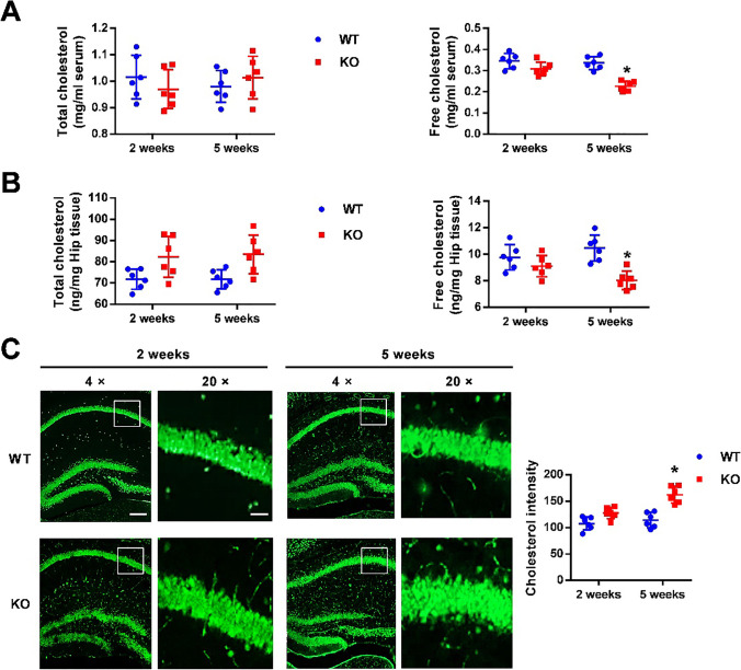
Fig. 6.
Excessive cholesterol is accumulated in the adolescent Fmr1 KO hippocampus. A, B Levels of total cholesterol and free cholesterol were detected in serum (A) and hippocampus tissue (B) from WT and KO mice. Free cholesterol was reduced over 5 weeks in KO mice. n = 6 mice per group; * p < 0.05 versus WT. Cholesterol content was normalized with protein and expressed as ng/mg of tissue. n = 6 mice per group; * p < 0.05 versus WT, two-way ANOVA with Tukey’s multiple comparisons test. C Filipin III staining showed the cholesterol (green) in the hippocampal CA1 region under 4 × and 20 × magnification, respectively. Green represents positive neurons. The statistical results showed that the cholesterol intensity in CA1 region increased in 5-week-old KO mice. Left: scale bar = 1000 μm; Right: scale bar = 100 μm. n = 6 mice per group; * p < 0.05 versus WT, two-way ANOVA with Tukey’s multiple comparisons test

