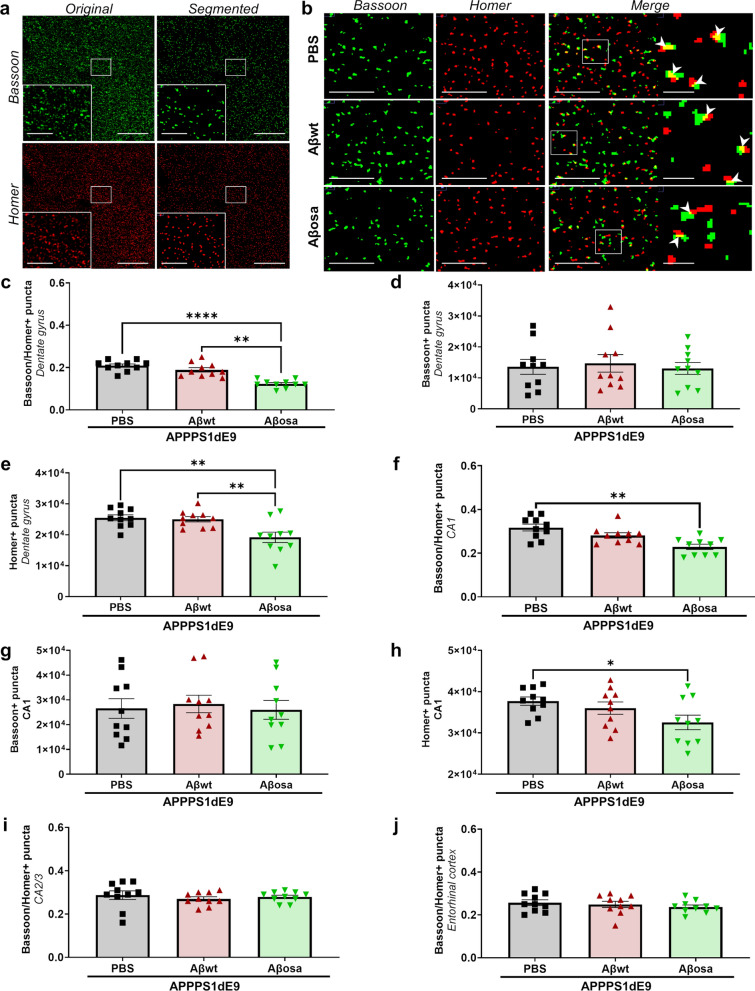
Fig. 5.
Aβosa exacerbates long-term synaptotoxicity in vivo. a. Representative views of original Bassoon/Homer images and segmented puncta in APPswe/PS1dE9 mice. Scale bars main images: 20 µm; Insets: 5 µm. b. Co-localisation puncta of Bassoon/Homer labels (white arrow). Scale bars main images: 5 µm; Insets: 1 µm. c-h. Quantification of synaptic density from Bassoon/Homer colocalization (c, f), Bassoon (d, g) and Homer (e, h) in the dentate gyrus and and CA1 showed decrease of synaptic density and post-synaptic density (Homer) in the dentate gyrus and CA1 of Aβosa-inoculated APPswe/PS1dE9 mice (c: Bassoon/Homer in dentate gyrus—overall effect: p < 0.0001 (Kruskal–Wallis). Post-hoc evaluation with Dunn’s multiple comparisons: Aβosa- versus PBS-inoculated APPswe/PS1dE9: p < 0.0001; Aβwt- versus Aβosa-inoculated APPswe/PS1dE9: p = 0.003; e: Homer in dentate gyrus—overall effect: p = 0.0015 (Kruskal–Wallis). Post-hoc evaluation with Dunn’s multiple comparisons: Aβosa- versus PBS-inoculated APPswe/PS1dE9: p = 0.0029; Aβwt- versus Aβosa-inoculated APPswe/PS1dE9: p = 0.0058; f: Bassoon/Homer in CA1—overall effect: p = 0.002 (Kruskal–Wallis). Post-hoc evaluation with Dunn’s multiple comparisons: Aβosa- versus PBS-inoculated animals: p = 0.002; h: Homer in CA1—overall effect: p = 0.0535 (Kruskal–Wallis). Post-hoc evaluation with Dunn’s multiple comparisons: Aβosa- versus PBS-inoculated APPswe/PS1dE9: p = 0.0469. There were no changes in the different groups in the CA2/3 (i) and in the entorhinal cortex (j). nAPP/PS1-PBS = 10, nAβwt = 10, nAβosa = 10 mice. For each image, quantification was made on 26 sections separated by 0.2 µm step. A surface of 81.92*81.92µm2 was measured for each section. Data are shown as mean ± s.e.m

