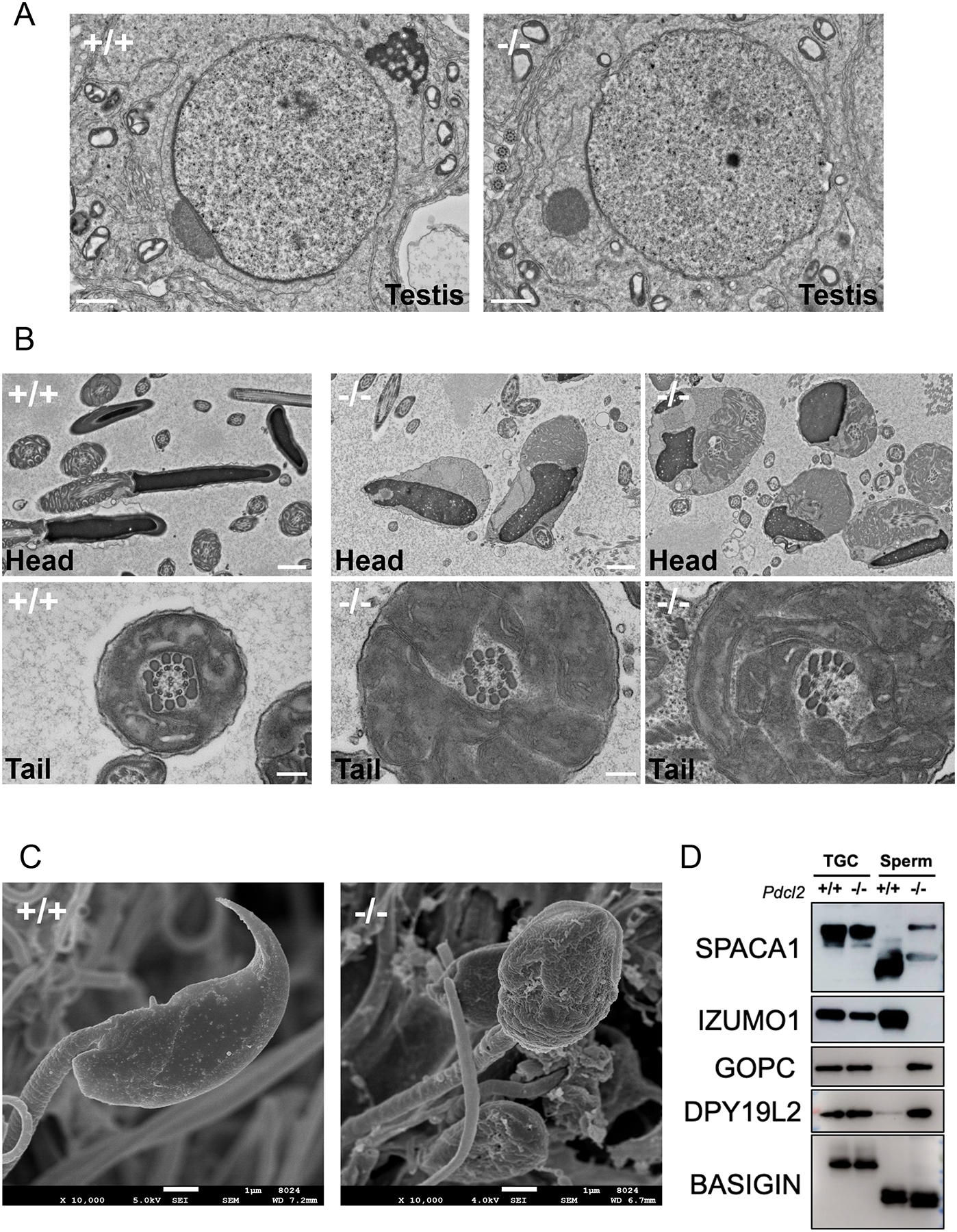
Figure 3: Impaired sperm acrosome formation in Pdcl2−/− male mice.

(A) TEM observation of round spermatids. Abnormal dissociation of the acrosome and the granule during cap biogenesis was observed in step 7 of round spermatid. Scale bars: 1 μm.
(B) TEM observation of mature spermatozoa in the cauda epididymis. Pdcl2 null sperm heads have misshapen nuclei and disorganized mitochondria in cytoplasmic droplets. There was no evidence of an acrosome cap. Pdcl2 null spermatozoa have disrupted axonemes and incomplete midpieces. Scale bars: 1 μm.
(C) SEM observation of mature spermatozoa extracted from the cauda epididymis. Scale bars: 1 μm.
(D) Immunoblot analysis of proteins related to acrosome biogenesis in wild-type and Pdcl2−/− testicular germ cells and cauda epididymal spermatozoa: SPACA1, IZUMO1, GOPC, DPY19L2. BASIGIN was used as a loading control.
