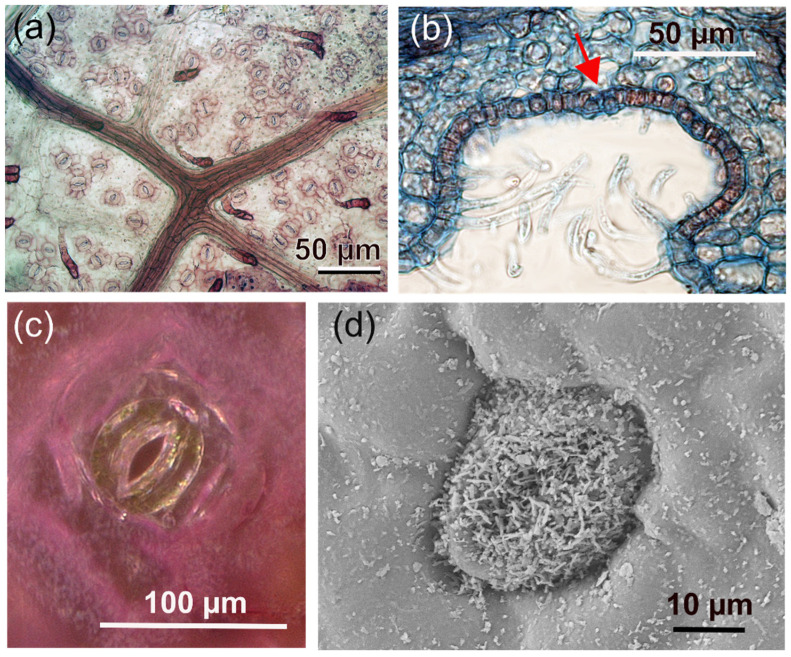
Figure 4.
Examples of stomata on leaves. (a) Close-up of a leaf of Fagus sylvatica showing stomata distributed over the leaf surface. The stomata resemble coffee beans and consist of two guard cells that are aligned parallel to each other and form the stomatal aperture between them. The dark branched structure is a part of the leaf venation. (b) A cross-section through a stomatal crypt of Nerium oleander. The red arrow indicates a stoma. (c) A stoma of Tradescantia pallida. Due to pigments, the epidermis cells and subsidiary cells are reddish, whereas the two guard cells of the stomata apparatus are green, indicating the presence of chloroplasts (which is characteristic for guard cells). (d) Scanning electron microscopic (SEM) image of a stoma of Laurus nobilis (laurel) decorated with wax crystal structures.

