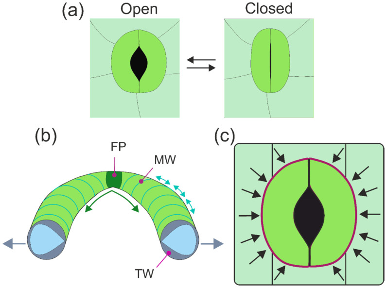
Figure 5.
The opening mechanism of stomata. (a) In many stomatal types, the aperture changes by backward bending of the guard cells. (b) Illustration of the various discussed mechanisms of shape change of guard cells. Often, the cell wall on the ventral side (meaning the cell side facing the aperture) is thicker (TW) than on the dorsal side (meaning the cell side opposite to the aperture), illustrated by the “cut face” of the picture (cell wall material: greyish; cell lumen: light blue). MW: micellar radial arrangement of cellulose bundles, as indicated by the light turquoise lines. FP: fixed polar ends of the guard cells. For more details and explanations of the opening mechanism, see text. (c) In many stomatal types, the shape change of guard cells can also be affected by adjacent epidermis cells (subsidiary cells), depicted here in simplified manner as rectangles, which are pressing against the neighboring walls of the guard cells (indicated by vectors and the purple line).

