
Figure 6. Effects of CBD on the mRNA and protein levels of WT1 in TM4 and human Sertoli cells.
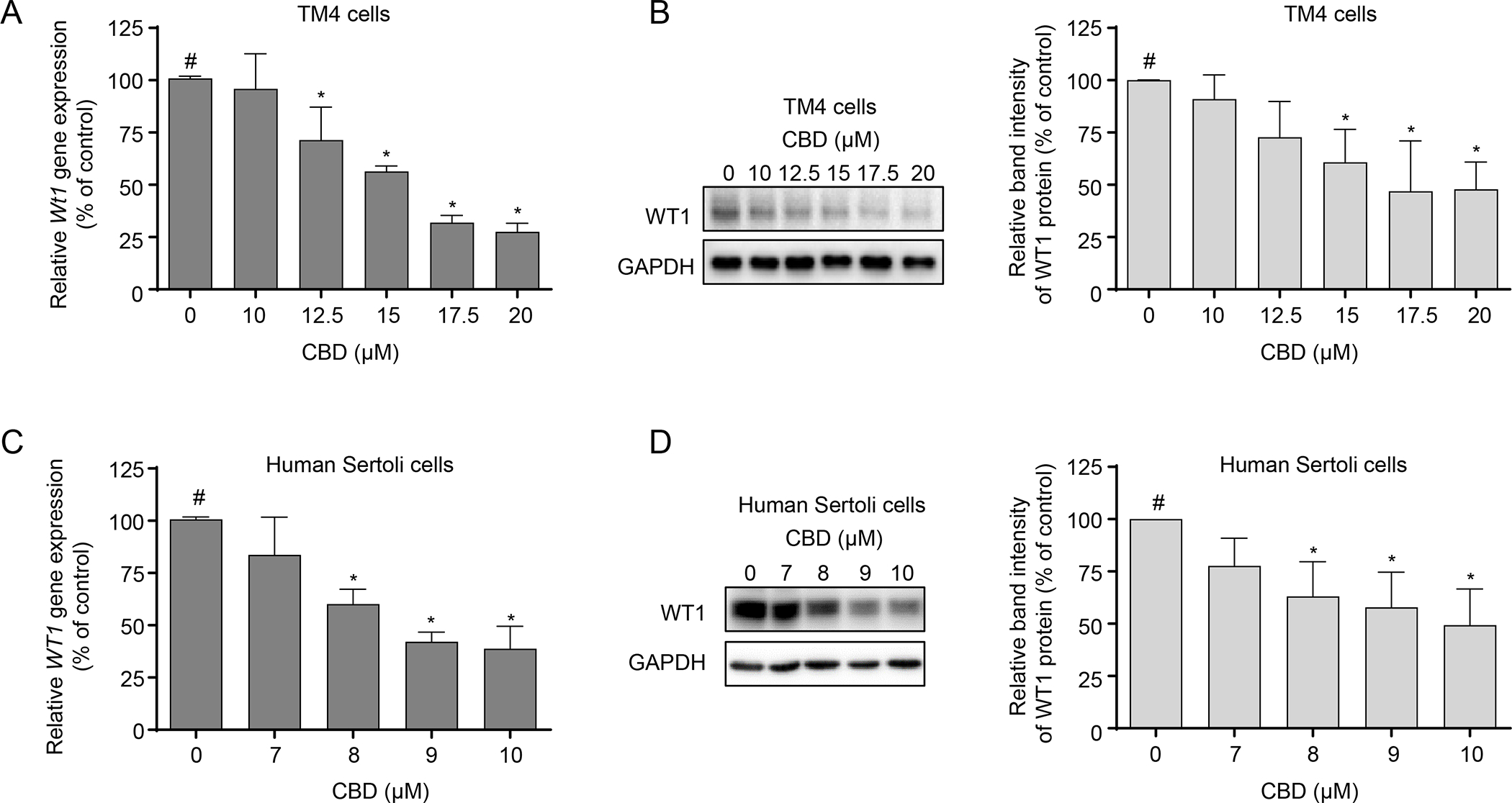
TM4 and human Sertoli cells were treated with DMSO (0 μM) or the indicated CBD concentrations for 24 h. Relative expression of Wt1 was determined using real-time PCR in CBD-treated TM4 (A) and human Sertoli cells (C). The results shown are means ± SD (n=3). #, significant concentration-related linear trend. *, significantly different from the DMSO control. Western blot analysis of WT1 was conducted in CBD-treated TM4 (B) and human Sertoli cells (D). Representative Western blots are shown in the left panel, and quantification is shown in the right panel. The results represent means ± SD (n=3). #, significant concentration-related linear trend. *, significantly different from the DMSO control.