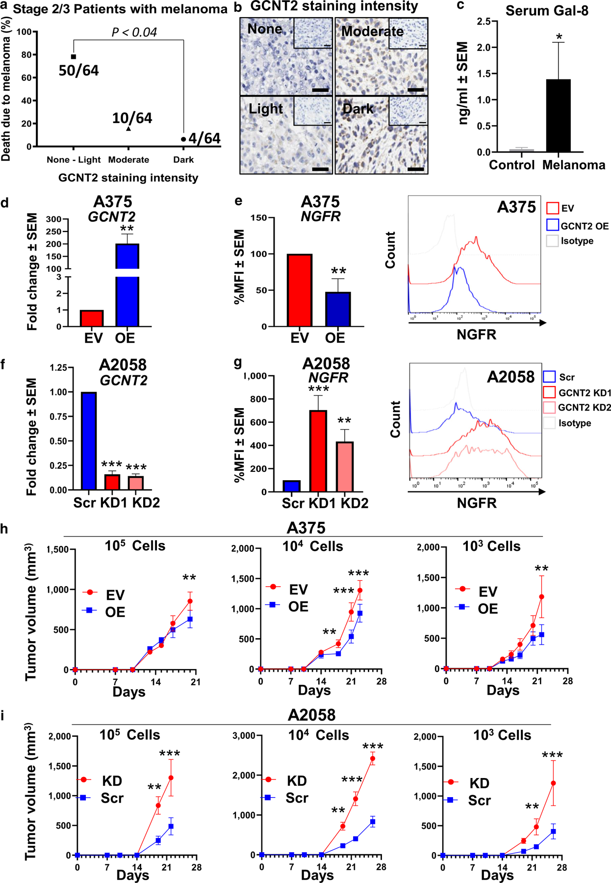
Figure 3. Patients with melanoma with a poor outcome had low GCNT2 levels, the sera from patients with melanoma were elevated for Gal-8, and GCNT2 loss increased the expression of TIC markers on MM cells.

(a) IHC analysis of GCNT2 expression in patient melanomas. GCNT2 expression score on melanoma (n = 64) (range = 0–3: 0 = no staining, 1 = light staining, 2 = moderate staining, and 3 = dark staining) plotted against patient mortality outcome. (b) Representative IHC image. (c) ELISA of Gal-8 was performed on sera from normal healthy volunteers (n = 5) and patients with MM (n = 13). RT-qPCR analysis of GCNT2 in GCNT2-OE and -KD (d) A375 and (f) A2058 cells. Flow cytometry of NGFR on (e) GCNT2 EV and OE A375 cells and on (g) GCNT2 Scr and KD A2058 cells. In vivo limiting dilution assay of (h) A375 EV/OE cells and (i) A2058 Src/KD using cell numbers ranging from 105 to 103. At least four biological replicates were performed. ***P < 0.001, **P < 0.01, and *P < 0.05. EV, empty vector; Gal-8, galectin 8; GCNT2, β1,6 N-acetylglucosaminyltransferase 2; IHC, immunohistochemistry; KD, knockdown; MM, metastatic melanoma; OE, overexpressed; Scr, scrambled; TIC, tumor-initiating cell.
