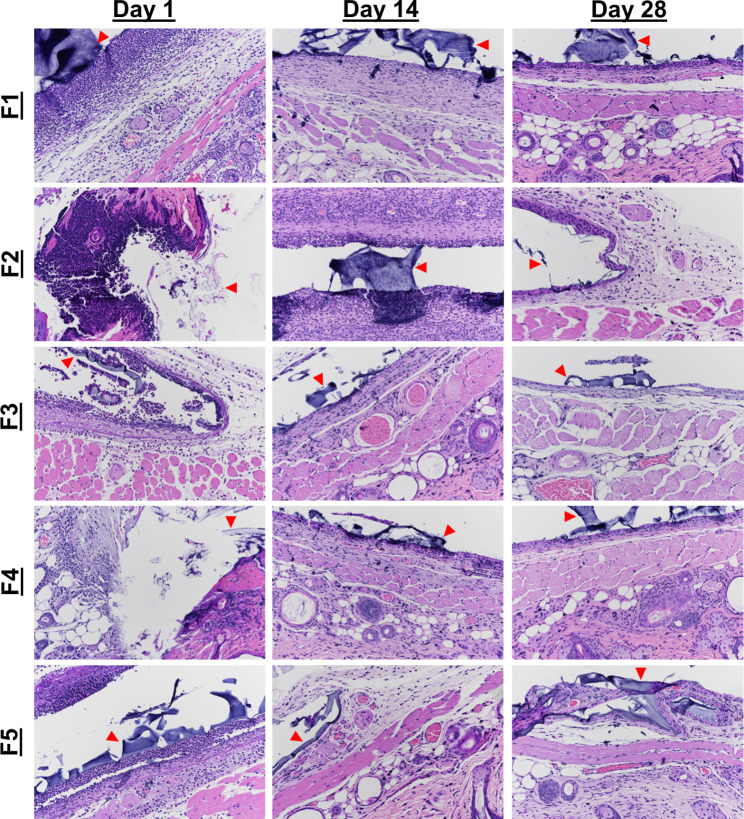
Fig. 4.
H&E stained tissue samples of SKH1-E taken from implant sites of hydrogels. Hydrogels of formulations 1–5 were explanted at days 1, 7, 14, and 28. The hydrogel itself or locations of the hydrogels are marked by arrows. In all formulations, we see heavy neutrophilic infiltration on day 1, with less on day 14, and resolution by day 28. The severity of acute inflammation is higher in formulations 1 and 2 compared to 4 and formulation 3 relative to formulation 5. Formulation 3, however, has fewer neutrophils than formulations 1 and 2. All formulations show an increase in edema, neovascularization, and fibrosis with time. Images were taken at 20x magnification, and gels are indicated by red arrows

