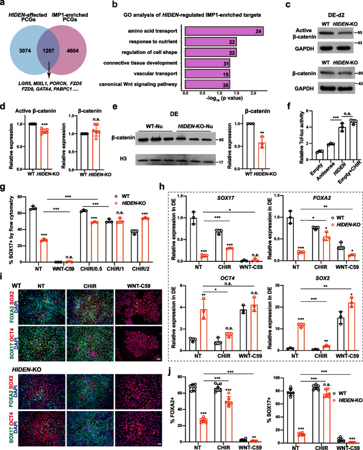
Fig. 5.
WNT signaling pathway acts as downstream of HIDEN/IMP1. a Venn diagram indicates the overlapped genes of differentially expressed PCGs upon HIDEN knockout in DE cells and IMP1-enriched targets identified by IMP1 RIP-seq in DE cells. b GO analysis of the overlapped genes from a. c, d HIDEN knockout led to reduced active β-catenin and unaltered total β-catenin level compared to wildtype after two days’ DE differentiation, as shown by Western blot (c) and statistical results (d) (n = 6). e The protein level of β-catenin in nuclear fraction of WT or HIDEN-KO DE cells (n = 3). f The TCF-luciferase activity in 293 T when transfected with HIDEN, antisense control or empty vector (n = 3). Cells treated with 1 μM CHIR-99021 were used as positive controls. g-j Flow cytometric analysis of SOX17-positive cells (g), the presentative endoderm genes expression revealed by RT-qPCR (h) and immunostaining (i-j) in wildtype or HIDEN-KO cells after manipulating WNT signaling through small molecular inhibitors during DE differentiation (n = 3). WNT signaling activator CHIR-99021, with different concentrations (0.5 μM, 1 μM and 2 μM) at (g) and 1 μM at (h-j), and WNT signaling inhibitor WNT-C59 (1 μM) were used. NT indicated for non-treated group. i Scale bar = 50 μm. j Quantitative results of SOX17- and FOXA2-positive cells were shown (n = 6)

