Fig. 6.
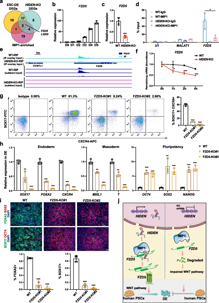
HIDEN-IMP1 stabilized FZD5 mRNA to contribute to DE differentiation. a Venn diagram indicated the overlapping of WNT-associated differentially expressed genes during DE differentiation (yellow circle), WNT-associated HIDEN-regulated genes (green circle), and IMP1-enriched WNT-associated genes (pink circle). The WNT-associated genes were listed in Additional file 8: Table S6 (from online WNT website: http://web.stanford.edu/group/nusselab/cgi-bin/wnt/). b The time course expression of FZD5 during DE differentiation, as shown by RT-qPCR (n = 3). c The relative expression of FZD5 in wildtype or HIDEN-KO DE cells was determined by RT-qPCR (n = 3). d RT-qPCR following IMP1 RIP was performed for determination of the indicated transcripts enrichment by IMP1 in wildtype or HIDEN-KO DE cells (n = 3). RNA levels were normalized to input. U1 and MALAT1 were considered as negative controls. e The visualization of IMP1 binding on FZD5 mRNA identified by IMP1 RIP-seq in wildtype or HIDEN-knockout DE cells. A detailed description was in Methods. f RNA stability assay of FZD5 in wildtype or HIDEN-KO DE cells treated with Actinomycin D (n = 3). g Flow cytometric analysis of SOX17+CXCR4+ cells in wildtype or FZD5-KO DE cells (n = 3). h RNA levels of representative endoderm genes, mesoderm genes and pluripotency genes in wildtype or FZD5-KO DE cells, determined by RT-qPCR (n = 3). i Immunofluorescent staining of DE markers (SOX17, FOXA2) and pluripotency markers (OCT4, SOX2) in wildtype or FZD5-KO DE cells. Quantitative results of SOX17- and FOXA2-positive cells were shown on the bottom (n = 5). Scale bar = 50 μm. j The functional model of HIDEN in human DE differentiation

