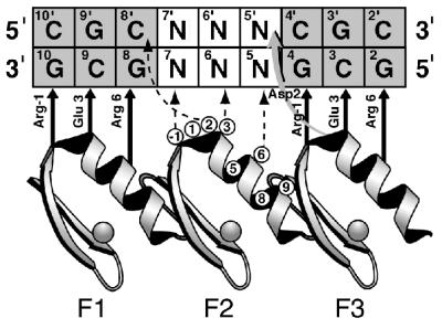
Figure 1.
Model depicting interactions between the Zif268 phage display library and the DNA used in microarray binding experiments. The three zinc fingers of Zif268 (F1, F2 and F3) are aligned to show contacts to the nucleotides of the DNA binding site as inferred from the crystal structure of Zif268 and biochemical experiments. The zinc finger amino acid positions are numbered relative to the first helical residue (position 1). The randomized positions in the α-helix of the second finger are circled. DNA base pairs marked N were fixed as particular sequences (11). © Copyright (2001) National Academy of Sciences of the USA.

