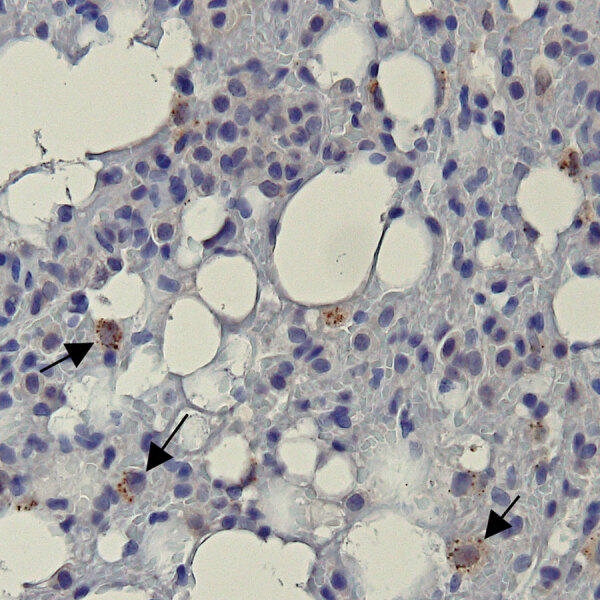
Figure 2.

Representative bat lung tissue showing Ebola virus (EBOV) cytoplasmic granules in study of histopathologic changes in Mops condylurus bats naturally infected with Bombali virus, Kenya. We labeled lung tissue sections by using rabbit polyclonal serum against EBOV matrix protein VP40 and detected antigen by using a chromogenic horse radish peroxidase substrate. The sections were then counterstained with hematoxylin. Arrow indicates granular cytoplasmic immunopositivity for EBOV VP40 antigen. Original magnification ×400. VP, viral protein.
