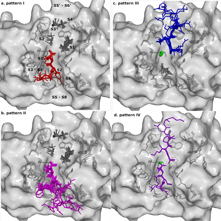
Fig. 5. Binding geometry of peptides.
Crystal structures of all complexes of cathepsin V were superimposed. Bound peptides are shown on the background of cathepsin V active site residues shown as gray sticks and the surface is shown in semi-transparent gray. The bound peptides are shown as stick models colored according to the patterns (a) I (red), (b) II (purple), (c) III (blue) and (d) IV (violet). Chlorine ions are shown as green balls and MPD molecules as dark gray sticks. The figures were generated with MAIN59 and rendered with RASTER 3D66.

