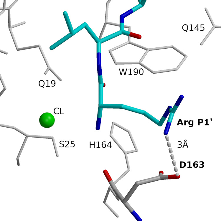
Fig. 6. Binding specificity between Arg at P1′ and D163 (cathepsin V).

Arg and Leu of peptide RLSAKP (non-protected), bound at subsites S1′ and S2′ are shown in the bond model in blue (nitrogen), red (oxygen), and cyan (carbon). D163 is shown in the bond model in blue (nitrogen), red (oxygen), and gray (carbon). Hydrogen bond is shown with a dashed line. Neighboring residues and the chlorine ion are also provided. Figures in the panel were prepared using MAIN59 and rendered using Raster 3D66.
