
Figure 3.
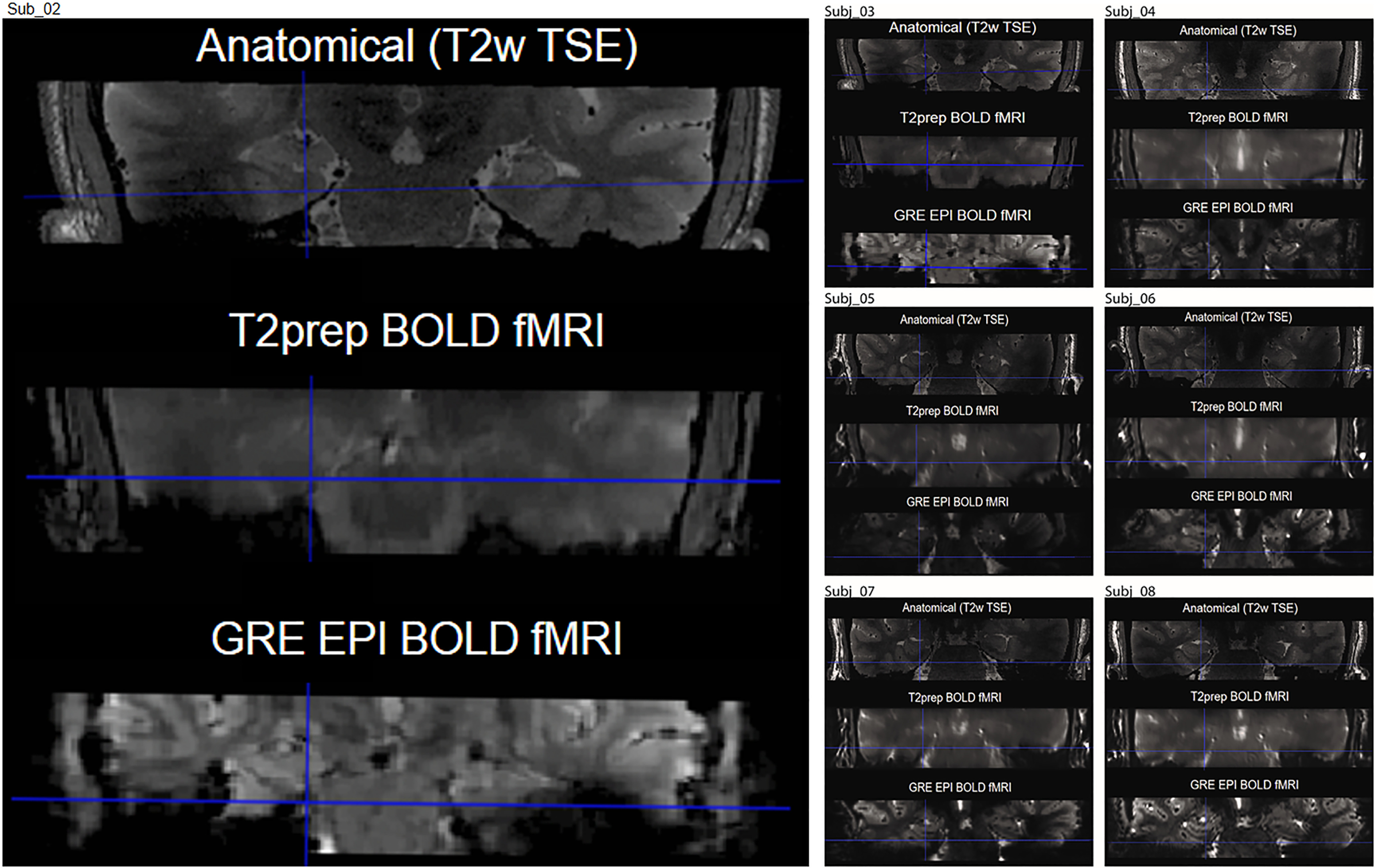
Representative images acquired using GRE EPI BOLD and T2prep BOLD fMRI (coronal view). In most subjects, susceptibility artifacts (signal dropout and geometric distortion caused by the nearby cavity in the ear canal) in the entorhinal cortex were significantly reduced in T2prep BOLD (middle) compared with GRE EPI BOLD (bottom). The high resolution T2-weighted image is shown as a dropout and distortion free anatomic reference (top).