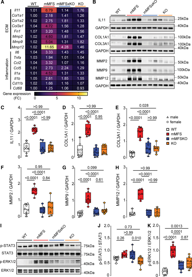
Figure 4.
Lack of IL11 (interleukin-11) signaling in mouse model of Marfan Syndrome (mMFS) mice reduces pulmonary extracellular matrix modification and inflammation. A, Heat map representation of mean gene expression, as determined by real-time polymerase chain reaction, for ECM (extracellular matrix) genes (Il11, Col1a1, Col3a1, Fn1, Mmp2, Mmp9, and Mmp12) and inflammatory genes (Il6, Tnfa, Ccl2, Ccl5, Il1a, Il1b, Cd11b, and Cd68) in lung lysates of 16-week-old WT, mMFS, mMFSxKO, and KO mice (n=6). Data are expressed as relative fold-change to controls with colour intensity indicating fold-change. Quantitative data are presented in Table S1. Representative immunoblots (B) and densitometric analyses of WT, mMFS, mMFSxKO, and KO lungs probed for IL11 (C), COL1A1 (D), COL3A1 (E), MMP2 (F), MMP9 (G), and MMP12 (H) normalized to GAPDH expression (MMP12: n=3M, 3F; others: n=4M, 4F). Representative immunoblots (I) and densitometry of WT, mMFS, mMFSxKO, and KO lungs probed for phosphorylated (p-) STAT3 (J) and ERK1/2 (K) normalized to their respective total expression (n=4M, 4F). Data shown are expressed as median±IQR; whiskers denote minimum and maximum values. Sexes are indicated as symbols for males (▲) and females (●). Statistical analysis was performed by 2-way ANOVA with Sidak multiple comparisons (C–H and J–K) and P values reported for the following groups: WT vs mMFS, mMFS vs mMFSxKO, mMFSxKO vs KO, and WT vs KO, respectively.

