Figure 2.
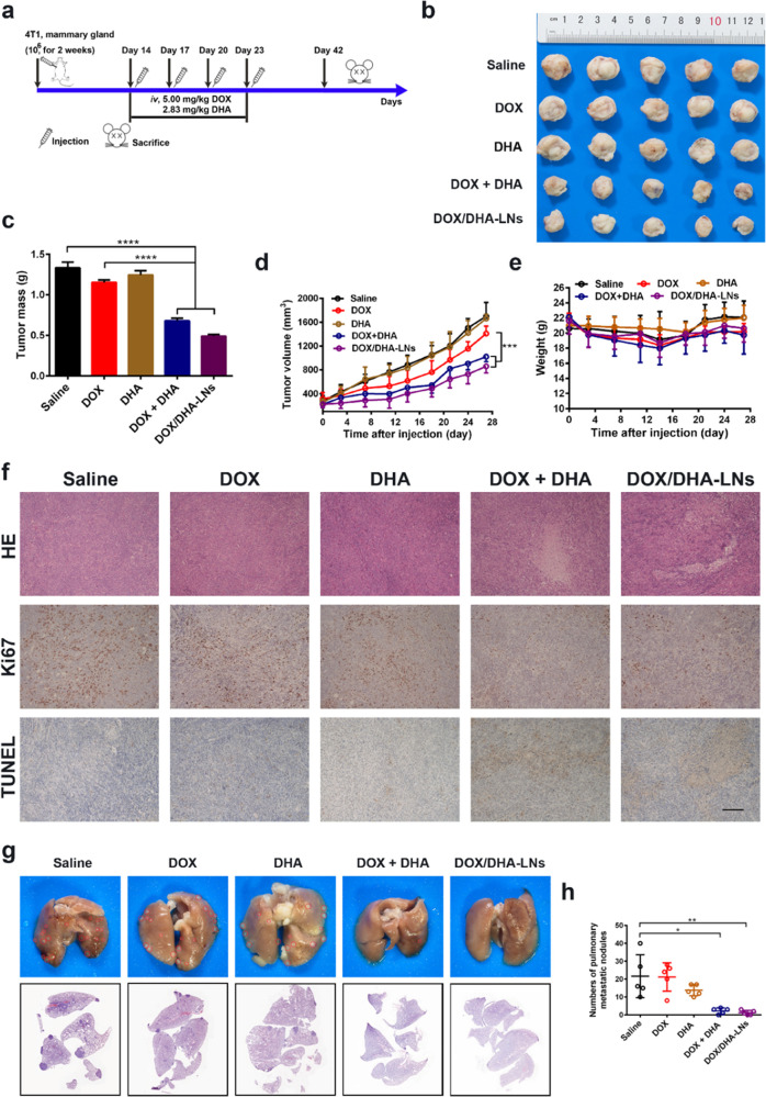
Tumor inhibitory effect and antimetastasis effect in vivo. (a) Design and timeline of animal experiment to test antimetastasis and survival in vivo. iv., intravenous. (b) Photographs of tumors. (c) The tumor weights at the end of the experiment. (d) The tumor volumes after treating with saline, DOX, DHA, DOX + DHA, and DOX/DHA-LNs for four times with equivalent doses of 5.00 mg kg–1 of DOX and 2.83 mg kg–1 of DHA. (e) The variation profiles of body weights. (f) Representative images of tumor sections stained for hematoxylin and eosin (H&E), Ki67, and TUNEL. Scale bar = 200 μm. Data represent mean ± SD (n = 5). (g) Representative images and histopathologic examination of lungs from 4T1 tumor-bearing mice after treating with saline, DOX, DHA, DOX + DHA, and DOX/DHA-LNs for four times with 5.00 mg kg–1 of DOX and 2.83 mg kg–1 of DHA. (h) Quantitative analysis of the pulmonary metastatic nodules of the lungs. Data represent mean ± SD (n = 5). P values were determined by one-way ANOVA with post hoc Tukey test and represented using *P < 0.05, **P < 0.01, ***P < 0.001, and ****P < 0.0001.

