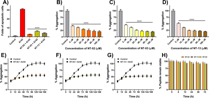
Figure 4.
(A) Quantification of percentage apoptotic cells of different treatments shows the significant rescue of cells from Aβ42-induced apoptosis by NT-02, NT-03, and NT-13 peptides. Effects of NT peptides on Aβ42 aggregation measured by the ThT fluorescence assay. (B) Peptides NT-02, (C) NT-03, and (D) NT-13 in Aβ42 aggregation. The control sample represents the Aβ42 peptide (5 μM) alone. (E–G) Effects of peptides NT-02, NT-03, and NT-13 on Aβ42 aggregation for 7 days. Aβ42 (5 μM) and peptide at a 10 μM concentration. Control sample represents the Aβ42 peptide (5 μM) alone. (H) Serum stability of NT peptides in human serum up to 24 h. Error bars represent mean ± standard deviation (SD), n = 3. Statistical data were analyzed by a one-way ANOVA test by the multiple comparison tests (*p < 0.05, **p < 0.01, ***p < 0.001, ****p < 0.0001, vs Aβ42) using software GraphPad Prism (ISI, San Diego, CA).

