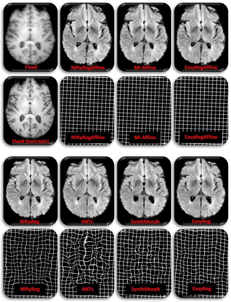
Figure 5.
Sample registration from SagT1–AxFLAIR setup, i.e., 5 mm axial FLAIR from ADNI, to 5 mm sagittal T1-weighted scan from IXI. Top left: axial slice of the fixed image and corresponding isotropic slice; the later was not used in registration; we display it for easier qualitative assessment of registration accuracy. Rest: corresponding registered slice and deformation field (represented as a deformed grid) for the seven competing methods.

