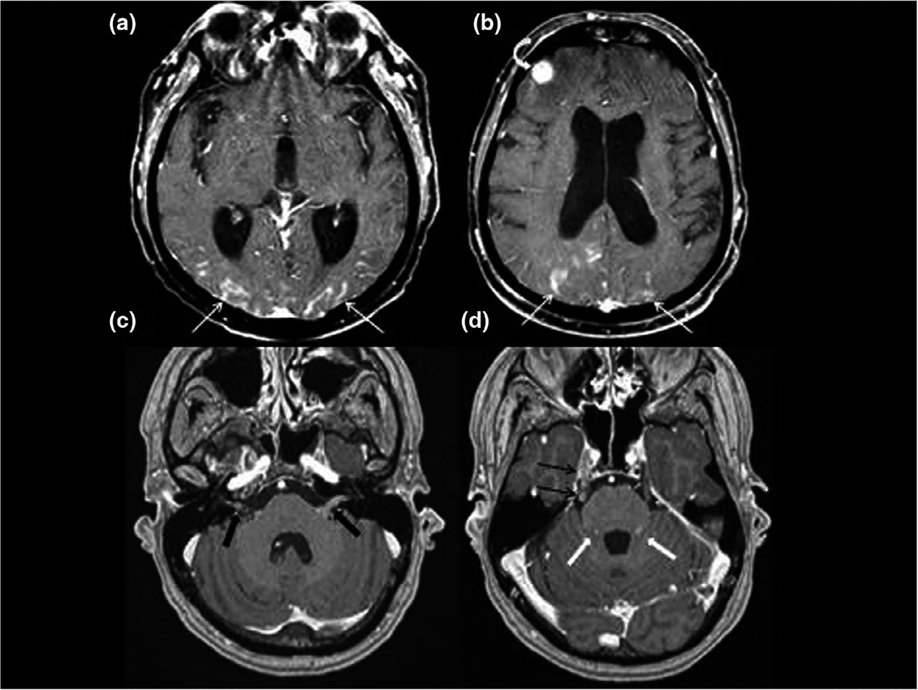
FIGURE 2.

Contrast enhanced axial imaging of the supratentorial brain (a,b) demonstrates curvilinear enhancement of the pial surface of the brain from leptomeningeal disease (LMD). Extensive pial enhancement involves the occipital and parietal cortex bilaterally (white arrows) as well as a focal nodular deposit involving the right frontal cortex (white curved arrow). Contrast enhanced axial imaging of the posterior fossa (c,d) demonstrates nodular enhancing LMD involving cranial nerves VII and VIII within the IAC and CPA (black block arrows), right cranial nerve V in lateral pontine cistern and Meckel’s cave (black arrows), as well as the folia of the cerebellum (white block arrows)
