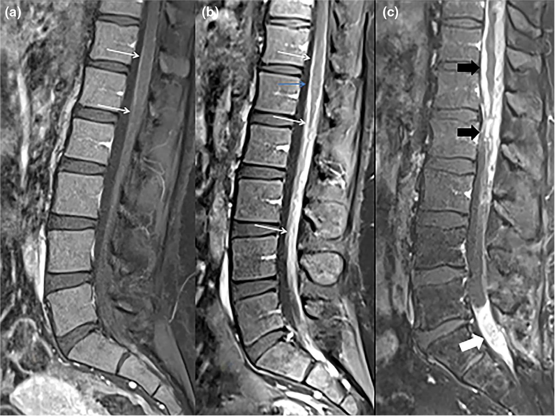
FIGURE 3.

Contrast enhanced sagittal images of three different patients. First patient (a) does not have leptomeningeal disease (LMD) and demonstrates normal minimal enhancement involving the pial surface of the lower thoracic cord and conus (white arrows). Patient b has extensive smooth LMD involving the conus and cauda equine (white arrows). Patient c has more extensive and nodular LMD involving the lower thoracic cord and conus (black block arrows) and extensive focal leptomeningeal disease involving the sacral nerve roots (white block arrow)
