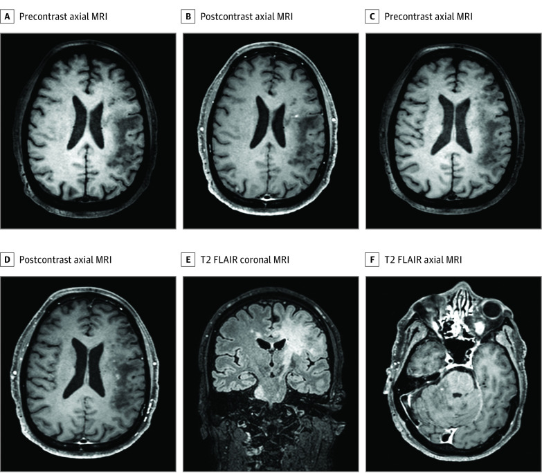
Figure 2. Enhancing Lesions on Brain Magnetic Resonance Imaging (MRI) of a Male Patient in the Sixth Decade of Life With Progressive Multifocal Leukoencephalopathy in the Context of Sarcoidosis (S-PML).
Brain MRI with corresponding T1 precontrast and postcontrast axial images and T2 fluid-attenuated inversion recovery (FLAIR) coronal and axial images of a male patient in the sixth decade of life who developed PML 7 years after pulmonary sarcoidosis. Punctate enhancement on a milky-way background is seen in panels B and D.

