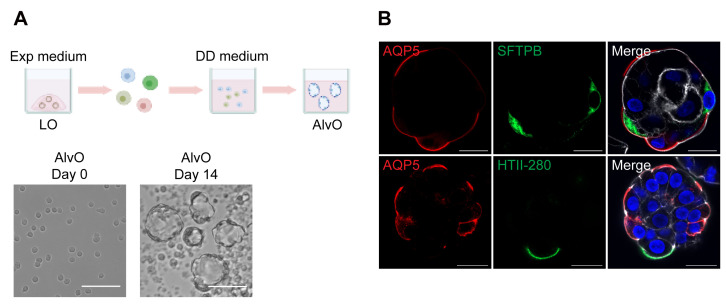
Figure 2. Generation of alveolar organoids.
(A). Top, a schematic graph outlines the distal differentiation protocol to generate alveolar organoids (AlvO) in distal differentiation medium (DD medium). Exp medium = expansion medium. Bottom, photomicrographs show single cell suspension on day 0 and mature alveolar organoids on day 14. Scale bar = 100 μm. (B). Alveolar organoids were applied to immunofluorescence staining to label AQP5+ AT1 cells (red) and SFTPB+/HTII-280+ AT2 cells (green). Nuclei and actin filaments were counterstained with DAPI (blue) and Phalloidin-647 (white), respectively. Scale bar = 20 μm.

