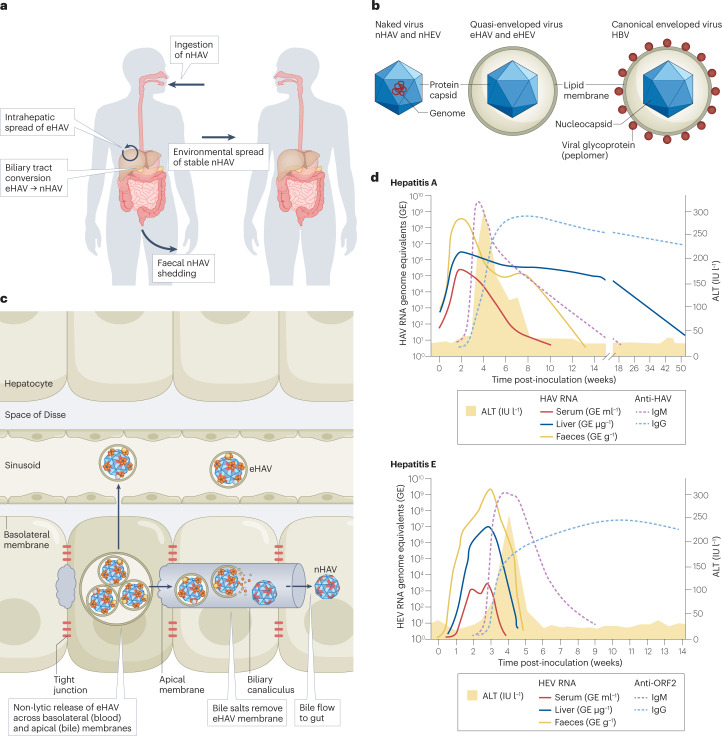
Fig. 1. Pathogenesis of enterically transmitted hepatitis A and hepatitis E virus.
a, Hepatitis A virus (HAV) life cycle, showing per-oral infection with naked HAV (nHAV), in-host spread of quasi-enveloped HAV (eHAV) and faecal shedding of nHAV resulting in environmental transmission to naive hosts. nHAV particles shed in faeces are produced by bile acid conversion of eHAV released from hepatocytes. The hepatitis E virus (HEV) life cycle is similar (not shown). b, Basic structures of naked versus quasi-enveloped hepatitis viruses and a canonical enveloped virus (hepatitis B virus (HBV)), showing the absence of virus-encoded proteins on the surface of quasi-enveloped virions11,31. c, Liver architecture, showing basolateral release of eHAV from polarized hepatocytes into blood flowing through hepatic sinusoids and apical release into a bile canaliculus. The quasi-envelope is stripped from eHAV by bile acids, resulting in faecal shedding of nHAV24. Events are similar in hepatitis E (not shown). d, Virological and serological markers in acute hepatitis A (top) and hepatitis E (bottom). ALT, alanine aminotransferase; IgG, immunoglobulin G; IgM, immunoglobulin M; IU l−1, international units per litre.

