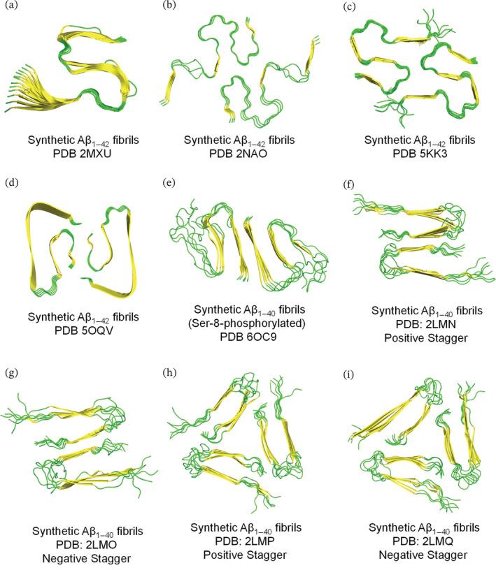
FIGURE 2.

Resolved ssNMR/Cryo‐EM structures of synthetically prepared Aβ1–42/Aβ1–40 fibrils. Top view representation was used to demonstrate the fibril symmetry (i.e., conformation and number of molecules per fibril layer). (a–i) Fibrillar models are generated on PyMol using the protein data bank entries and are colored based on secondary structure (yellow for β‐sheets and green for loops). The terms, positive and negative stagger, in panels f–i describe the conformation of Aβ1–40 which spans two different z‐planes implying that each fibril layer is not occupied by a single Aβ1–40 molecule.
