Figure 2:
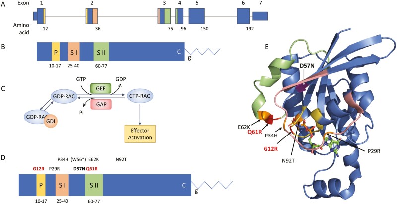
RAC2 gene and protein. (A) Genomic schematic for RAC2. Exons are numbered above; the number of the last amino acid codon in each exon is shown below. Exon 7 is untranslated. (B) Linear protein cartoon of RAC2 showing locations of specific domains. P, P-loop (yellow); S I, Switch I domain (peach); S II, Switch II domain (green); C, CXXL prenylation motif; g, geranylgeranyl moiety. (C) RAC2 bound to GDI is unable to interact with effectors. Release from GDI and interaction with GEFs removes GDP allowing binding of GTP. RAC2-GTP can then interact with downstream effectors. RAC2-GAP interactions lead to hydrolysis of GTP. (D) Reported RAC2 mutations shown on linear RAC2 protein with domains noted. Mutations in bold are constitutively active (red) or inactive (black). Mutation in parenthesis undergoes nonsense-mediated decay and does not result in a protein product. The remaining activating mutations maintain intrinsic GTP hydrolysis. (E) Three-dimensional structure of the related RAC1 (3TH5) [39] showing domains colored as in B and location of mutations. GTP shown as sticks.

