FIGURE 2.

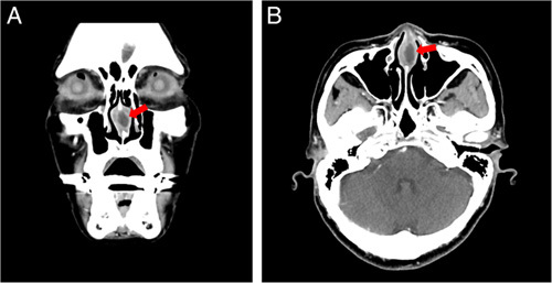
Coronal (A) and axial (B) views of the contrast-enhanced CT of the face revealed a low-density lesion measuring 19 mm×14 mm with rim enhancement inside the anterior nasal septum (red arrows).

Coronal (A) and axial (B) views of the contrast-enhanced CT of the face revealed a low-density lesion measuring 19 mm×14 mm with rim enhancement inside the anterior nasal septum (red arrows).