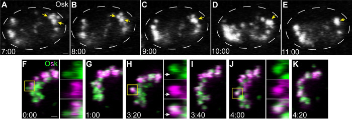
Fig 2. Germ granules grow by fusion in the pole cells.
Maximum intensity confocal z-projections of a 1 μm region of a representative pole cell at nc14 with endogenously tagged Osk-sfGFP (A–E) or Osk-Dendra2 (F–K). Osk-sfGFP and Osk-Dendra2 images were taken from a 5-min period of S1 Video and a 4-min period of S2 Video, respectively. Yellow arrows and boxes indicate germ granules that undergo fusion. Enlargements of the boxed regions in (F), (H), and (J), show the mixing of green and red (shown here in magenta) fluorescent Osk-Dendra2 signal over time. White arrows indicate a region of a granule where the magenta labeled and green labeled contents have yet not mixed after fusion. Time stamps indicate minutes:seconds. Scale bar: 1 μm.

