
Figure 2.
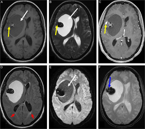
Axial brain MRI showing the right frontoparietal lesion measuring 72×53 mm in diameter exerting a midline shift estimated at 10 mm. This lesion had three different components. The cystic component had a hyposignal on T1-weighted sequences (A, white arrow) and hypersignal on T2-weighted sequences (B, white arrow) without restricted diffusion on diffusion-weighted sequences (E, white arrow). The solid component (mural nodule) had a heterogeneous signal on T1-weighted sequences (A, yellow arrow) and a frank hyposignal on T2-weighted sequences (B, yellow arrow) with major enhancement after chelates of gadolinium injection (C, yellow arrow). Note the third component, which was a calcium deposit having a signal void with a hyposignal on the gradient echo sequence (F, blue arrow). There was no peritumoral edema except a minor transependymal cerebrospinal fluid resorption at the level of the occipital horns on T2 fluid-attenuated inversion recovery sequences (D, red arrows).