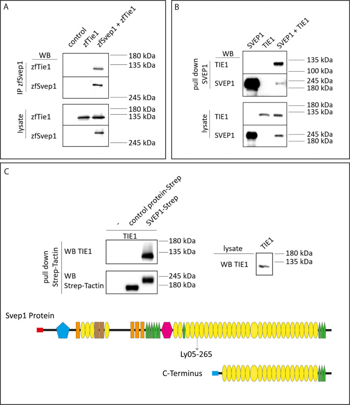
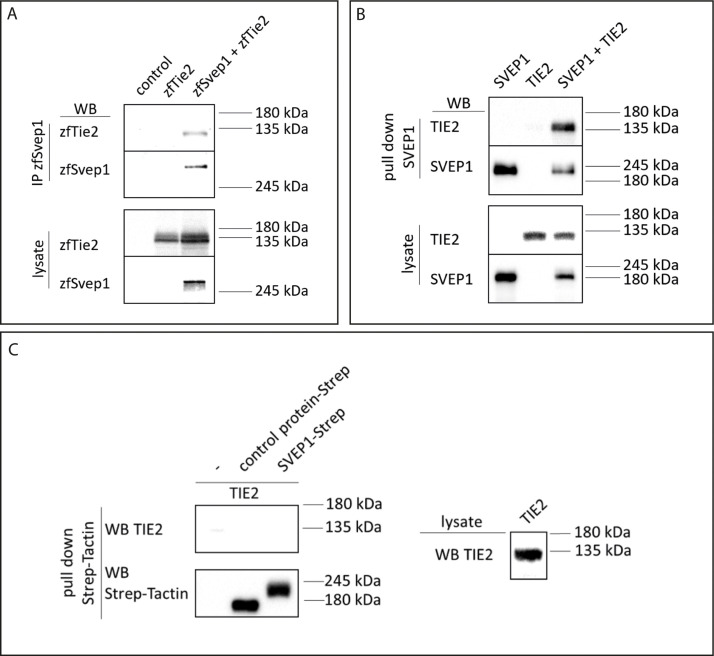
Figure 8. SVEP1 binds to TIE1.
(A) 293T HEK cells were transfected with zebrafish Svep1-HIS (zfSvep1) and zebrafish Tie1-HA (zfTie1). zfSvep1 was immunoprecipitated and associated Tie1 was detected by western blot. (B) Co-immunoprecipitation of C-terminal human SVEP1 co-transfected in 293T HEK cells with human TIE1. (C) Pull-down of recombinant C-terminal human SVEP1-Strep-tag II protein, which was incubated with TIE1 transfected 293T HEK cell lysates, shows binding of TIE1. Protein structure with all domains indicated and C-terminal part used for pull-down assays (adapted from Figure 2F of Karpanen et al., 2017, published under the CC BY-NC 4.0 license, https://creativecommons.org/licenses/by-nc/4.0/). It is not covered by the CC-BY 4.0 license and further reproduction of this panel would need to follow the terms of the CC BY-NC 4.0 license. Ly05-265 indicates position of stop codon in the zebrafish hu6985 allele (Karpanen et al., 2017), suggesting that the protein domains C-terminal to the nonsense allele are critical for function. Red and blue rectangle: signal peptide; blue pentagon: von Willebrand factor type A domain (vWF); orange rectangle: ephrin-receptor like domain; brown rectangle: Hyalin repeat; yellow ovals: SUSHI repeat; green pentagons: epidermal growth factor (EGF)-like and calcium-binding EGF-like domains; and pink hexagon: pentraxin domain (PTX).
© 2017, Karpanen et al
(C) Pull -down of recombinant C-terminal human SVEP1-Strep-tag II protein, which was incubated with TIE1 transfected 293 T HEK cell lysates, shows binding of TIE1. Protein structure with all domains indicated and C-terminal part used for pull down assays (adapted from Figure 2F of Karpanen et al., 2017, published under the CC BY-NC 4.0 license (https://creativecommons.org/licenses/by-nc/4.0/). It is not covered by the CC-BY 4.0 license and further reproduction of this panel would need to follow the terms of the CC BY-NC 4.0 license)


