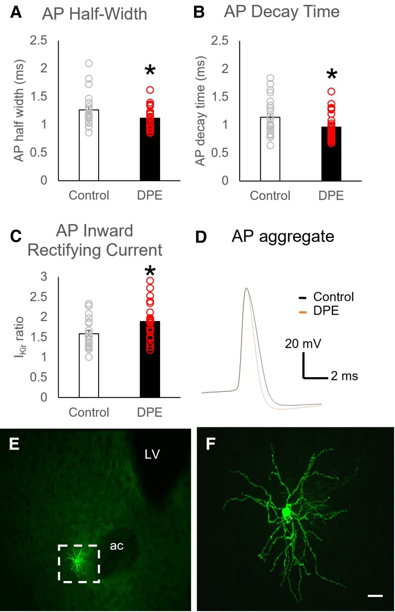
Fig. 3.
Changes in ventral striatum MSN properties in DPE mice. A) Action potential duration in MSNs, as measured by half-width, was reduced in DPE neurons (N = 22 neurons from 7 mice) relative to controls (N = 26 neurons from 6 mice), an effect driven by B) a faster action potential decay time. C) Inward-rectifying current was increased in MSNs of DPE neurons. D) Aggregate representation of MSN action potentials. E, F) Representative images show E) the morphology of a medium spiny neuron and F) the location of one recording site (site 4) in the ventral striatum. Scale bar is 20 µm. Error bars are SEM. *P < 0.05 by t test. ac, anterior commissure; LV, lateral ventricle.

