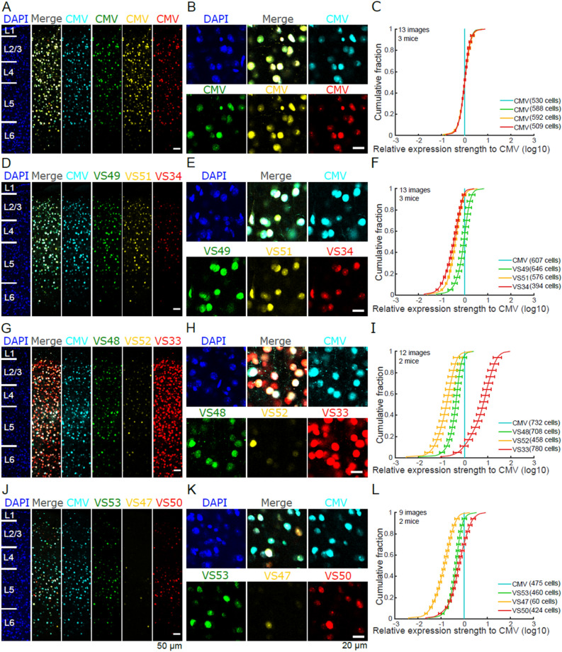
Figure 4.
Multiplexed fluorescence microscopy of nine validation sequences in coronal sections of the auditory cortex. (A) Positive control with all fluorophore variants (mTFP, EGFP, Venus, and tagRFP) driven by CMV promoter. Six neocortical layers indicated on DAPI image. (B) Zoomed in view of example region. (C) Cumulative distribution across cells of the relative fluorescence intensity of EGFP, Venus, and tagRFP compared to mTFP (relative expression strength) for the CMV-driven control constructs (mean across images ± SD). (D–L) Analogous to (A–C), but for the nine validation sequences examined, divided into three sets of three sequences (D–F, G–I, J–L). Each set also contains a CMV promoter-driven mTFP reporter, to which all the others are compared (F, I, L). VS33 drives high expression levels (G, H, I).

