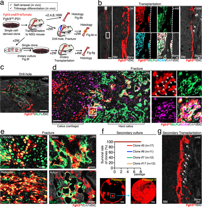
Fig. 6. Self-renewal and trilineage potential of Fgfr3+ endosteal stem cells in transplantation.
a Diagram of transplantation experiments of ex vivo expanded single cell-derived Fgfr3+ endosteal stem cells, involving NSG recipient mice, subsequent surgeries, secondary CFU-F, and transplantation assays. b NSG recipient femur transplanted with Fgfr3-creER+ endosteal stem cells after 2 weeks (left), 4 weeks with ALPL and ACAN staining (center), and 16 weeks (right). Scale bar: 500 µm (left), 20 µm (center), 40 µm (right). n = 4 clones. c Drill-hole injury of NSG recipient mice transplanted with Fgfr3-creER+ stem cells, at 2 weeks after surgery with ALPL staining. Scale bar: 200 µm. n = 4 clones. d, e Complete fracture of NSG recipient mice transplanted with Fgfr3-creER+ stem cells, at 2 weeks after fracture with ALPL and ACAN staining (d, e), with LepR and PLIN staining (e). Arrows: tdTomato, ALPL, and ACAN triple-positive cells. Scale bar: 100 µm (d), 20 µm (e). n = 4 clones. f Secondary CFU-F assays. The survival rate of individual tdTomato+ clones harvested from NSG recipient mice with Fgfr3-creER+ stem cell transplantation (top). Representative images of passages 0 and 8 (bottom). Scale bar: 5 mm. n = 4 clones (#5: n = 17, #6: n = 11, #7: n = 7, #17: n = 12,). g Secondary transplantation of Fgfr3-creER+ stem cells to NGS recipient femur, at 4 weeks after transplantation. Arrows: osteocytes derived from transplanted tdTomato+ stem cells. Scale bar: 20 µm. n = 4 clones.

