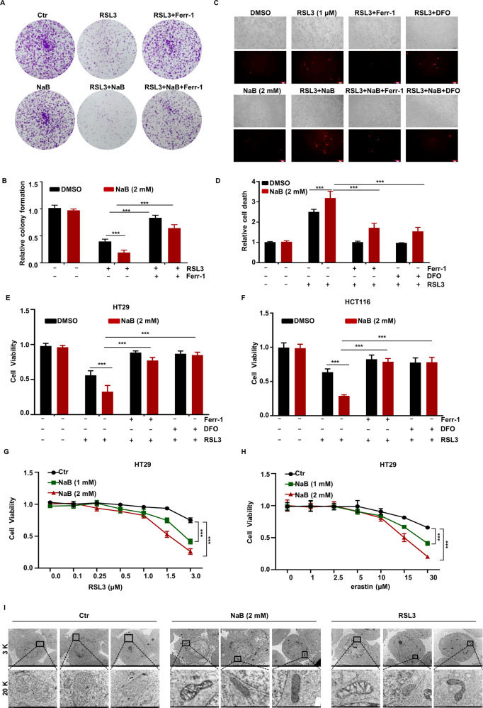
Fig. 1. Induction of cell death by butyrate in the presence of ferroptosis inducers.
HT29 cells were treated with NaB (2 mM) for 24 h in combination with RSL3 (1 μM) or Ferr-1 (5 μM), after which cell viability was detected by colony formation assay (A) and the quantitative data is presented (B). HT29 cells were treated with NaB (2 mM) for 24 h in combination with RSL3 (1 μM), Ferr-1 (5 μM), or DFO (100 μM) after which cell viability was detected by dead cell staining assay (C) and the quantitative data is presented (D). HT29 (E) or HCT116 (F) cells were treated with RSL3 (1 μM) for 6 h in combination with NaB (2 mM), Ferr-1 (5 μM) or DFO (100 μM), and the viability of indicated cells was examined using CCK-8. HT29 cells were treated with different concentrations of RSL3 for 6 h (G) or erastin for 20 h (H) in combination with indicated concentrations of NaB, and the cells viability was examined using CCK-8. I HT29 cells were treated with the RSL3 (1 μM) or NaB (2 mM) for 24 h, and analyze the ultrastructure of mitochondria with transmission electron microscopy.

