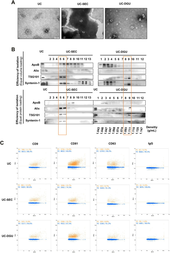
FIGURE 2.
Characterization of pEVs. (A) The morphological characteristics of pEVs were detected by TEM. (B) The EVs markers (Alix, TSG101 and Syntenin-1) and non-EVs marker (ApoB) were detected by Western blotting. 20 uL sample or 20 μg protein from each fraction was loaded. (C) The pEVs were fluorescently labeled with PE-conjugated EVs mAbs (CD9, CD63 and CD81), and the positive ratio was detected by nFCM measurement.

