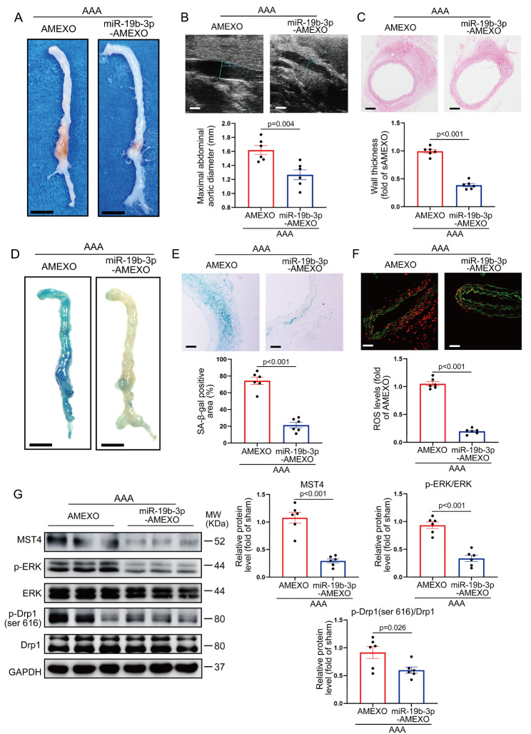
Fig. 8.
Overexpression of miR-19b-3p enhances the function of AMEXO in attenuating AAA formation and vascular senescence. (A) Representative photographs of the whole aorta from Ang II-induced AAA mice treated with AMEXO and miR-19b-3p-AMEXO. Scale bar: 2 mm. (B) Two-dimensional ultrasound imaging of abdominal aorta from Ang II-induced AAA mice treated with AMEXO and miR-19b-3p-AMEXO and analysis of the maximum aortic diameter (n = 6 independent experiments). Scale bar: 1 mm. (C) HE staining of abdominal aortic aneurysm sections from Ang II-induced AAA mice treated with AMEXO and miR-19b-3p-AMEXO and analysis of aortic thickness (n = 6 independent experiments). Scale bar: 150 μm. (D) Representative images showing SA-β-gal staining of the whole aorta from Ang II-induced AAA mice treated with AMEXO and miR-19b-3p-AMEXO. Scale bar: 2 mm. (E) Representative images of abdominal aortic sections after SA-β-gal staining from Ang II-induced AAA mice treated with AMEXO and miR-19b-3p-AMEXO. Percentage of SA-β-gal staining positive area was analyzed (n = 6 independent experiments). Scale bar: 50 μm. (F) DHE staining of abdominal aortic sections from Ang II-induced AAA mice treated with AMEXO and miR-19b-3p-AMEXO and ROS levels represented by DHE fluorescence intensity were analyzed. Red fluorescence indicates superoxide and green fluorescence indicates laminae (n = 6 independent experiments). Scale bar: 50 μm. (G) Western blotting analysis of the protein level of MST4, p-ERK and p-Drp1 (ser616) in the aorta from Ang II-induced AAA mice treated with AMEXO and miR-19b-3p-AMEXO (n = 6 independent experiments). Data are expressed as mean ± SEM. Two-tailed Student t test

