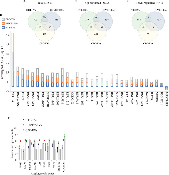
Figure 9.

Proangiogenic genes are secreted into extracellular vesicles (EVs) After lipid nanoparticle‐VEGF‐A (LNP‐VEGF‐A) treatment to cells. The Venn diagrams illustrate the overlapping DEGs (p < 0.05 and absolute log2 fold change > 1) in EVs from HTB cells (n = 4), HUVECs (n = 4), and CPCs (n = 4) after LNP treatment. A) The total overlap of DEGs in all three EV types. The overlaps of B) upregulated, and C) downregulated DEGs in all three EV types. D) A stacked bar plot showing log2 fold change of all the 32 DEGs from panels B and C, which demonstrate overlapping transcriptional patterns in the same direction (either up‐ or down‐regulated) in all EV types after LNP treatment. E) Top 10 angiogenesis genes identified in EVs of LNP‐treated cells. BiomaRt R package was applied, and 262 unique Ensembl gene IDs in EV‐mRNAs associated with angiogenesis (GO:0001525) were identified (a full list is provided in Tables S7–S9, Supporting Information); the top 10 angiogenic genes were selected (n = 4).
