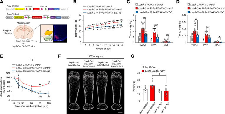
Figure 7. LepR-expressing VMH neurons contribute to LAT1-dependent regulation of systemic energy and bone homeostasis.
(A) Schematic diagram of the bilateral viral microinjection into the VMH and representative validation image of mCherry expression in the VMH. Scale bar, 500 μm. (B) Weekly body weight after injection of AAV-Control or AAV-Slc7a5 into the VMH is shown for LepR-Cre Slc7a5fl/fl mice and control mice (n = 8 to 12, **P < 0.01, ***P < 0.001: versus LepR-Cre/AAV-Control, #P < 0.05, ##P < 0.01, ###P < 0.001: versus LepR-Cre Slc7a5fl/fl/AAV-Control, 2-way ANOVA with Bonferroni post hoc test). (C and D) Adipose tissue weights (C) and adipose tissue weights normalized to body weight (D) are shown for LepR-Cre Slc7a5fl/fl mice and control mice injected with AAV-Control or AAV-Slc7a5 into the VMH at 16 weeks of age (n = 8, **P < 0.01, ***P < 0.001, #P < 0.05, ##P < 0.01, ###P < 0.001, 2-tailed Student’s t test with Bonferroni correction). (E) ITTs were performed in LepR-Cre Slc7a5fl/fl mice and control mice injected with AAV-Control or AAV-Slc7a5 into the VMH after a 6-hour fast at 16 weeks of age (n = 3 or 4, *P < 0.05, **P < 0.01: versus LepR-Cre/AAV-Control, #P < 0.05, ##P < 0.01, ###P < 0.001: versus LepR-Cre Slc7a5fl/fl/AAV-Control, 2-way ANOVA with Bonferroni post hoc test). (F) μCT analysis (scale bar, 1 mm) and (G) BV/TV ratio as determined by μCT of femurs from LepR-Cre Slc7a5fl/fl mice and control mice injected with AAV-Control or AAV-Slc7a5 into the VMH at 12–16 weeks of age (n = 6 to 11, *P < 0.05, #P < 0.05, 2-way ANOVA with Bonferroni post hoc test). All the mice used in this study were male.

