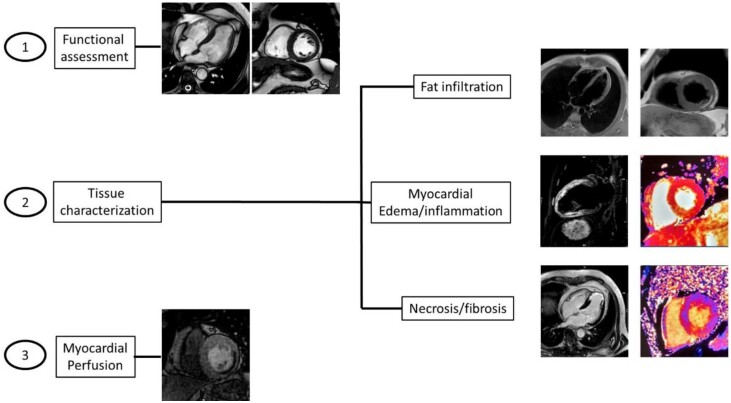
Figure 1.
The three-layered cardiac magnetic resonance approach in the characterization of ischemic cardiomyopathy: (1) Functional assessment: the images showed are cine steady-state free precession in four chamber and short-axis view. (2) Tissue characterization: T1 weighted images in four chamber and short-axis views for fat infiltration; cine steady-state free precession T2 weighted image in two chamber view and short-axis T2 mapping for myocardial edema/inflammation; late-gadolinium enhancement four chamber view and short-axis T1 mapping for necrosis/fibrosis assessment. (3) Myocardial perfusion: first-pass post-contrast short-axis image for the evaluation of perfusion defects.

