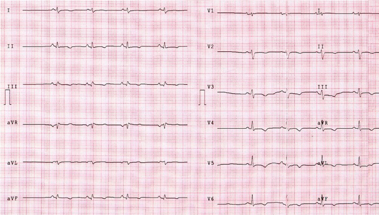
Figure 2.
ECG findings in a patient with ALVC. This 34-year-old man with history of ventricular arrhythmias had a CMR revealing biventricular dilation and a stria LGE pattern with subepicardial distribution, mainly involving the LV lateral wall. This patient showed the ‘classical’ low QRS voltages in limb leads and T-wave inversion in V2–V6 and inferior leads. CMR, cardiac magnetic resonance; LGE, late gadolinium enhancement; LV, left ventricular.

